What is Cyclic Peptide

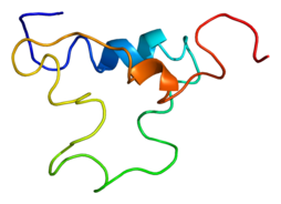
Cyclic peptides (or cyclic proteins) are polypeptide chains where in the amino termini and carboxyl termini, amino termini and side chain, carboxyl termini and side chain, or side chain and side chain are linked with a covalent bond that generates the ring. A number of cyclic peptides have been discovered in nature and a plethora have been synthesized in the laboratory. Their length ranges from just two amino acid residues to hundreds. These cyclic peptides have several applications in medicine and biology.

Cyclic peptides can be classified according to the types of bonds that comprise the ring.

Homodetic cyclic peptides, such as cyclosporine A, are those in which the ring is composed exclusively of normal peptide bonds (i.e. between the alpha carboxyl of one residue to the alpha amine of another). The smallest such species are 2,5-diketopiperazines, being derived from the cyclisaation of a dipeptide.

Cyclic isopeptides contain at least one non-alpha amide linkage, such as a linkage between the side chain of one residue to the alpha carboxyl group of another residue, as in microcystin and bacitracin.

Cyclic depsipeptides, such as aureobasidin A and HUN-7293, have at least one lactone (ester) linkage in place of one of the amides. Some cyclic depsipeptides are cyclized between the C-terminal carboxyl and the side chain of a Thr or Ser residue in the chain, such as kahalalide F, theonellapeptolide, and didemnin B.

Bicyclic peptides such as the amatoxins amanitin and phalloidin contain a bridging group, generally between two of the side chains. In the amatoxins, this bridge is formed as a thioether between the Trp and Cys residues. Other bicyclic peptides include echinomycin, triostin A, and Celogentin C. There are a number of cyclic peptide hormones which are cyclized through a disulfide bond between two cysteines, as in somatostatin and oxytocin.

One interesting property of cyclic peptides is that they tend to be extremely resistant to the process of digestion, enabling them to survive in the human digestive tract.[1] This trait makes cyclic peptides attractive to designers of protein-based drugs that may be used as scaffolds which, in theory, could be engineered to incorporate any arbitrary protein domain of medicinal value, in order to allow those components to be delivered orally. This is especially important for delivery of other proteins that would be destroyed without such implementation. Cyclic peptides are also “rigid” compared to the corresponding linear peptides, and this attribute promotes binding by removing the “entropic penalty”. For example, Daptomycin is a lipopeptide antibiotic used in the treatment of systemic and life-threatening infections caused by Gram-positive organisms. It is a naturally occurring compound found in the soil saprotroph Streptomyces roseosporus. Its distinct mechanism of action makes it useful in treating infections caused by multiple drug-resistant bacteria. It is marketed in the United States under the trade name Cubicin by Cubist Pharmaceuticals [2].

Karebay can synthetic cyclic peptide with Cyanine or other fluorescent moleculars , so that scientists can knew more details about its molecular mechanism.

Karebay (www.karebaybio.com) has a professional team devoted to peptide products synthesis and development. We offer high-quality biotechnology products for sale around the world, including over 1,000 catalog peptides, and nearly 100 pharmaceutical peptides and cosmetic peptides products.

Reference

[1] David J. Craik (17 March 2006). “Seamless Proteins Tie Up Their Loose Ends”. Science 311 (5767): 1563–7. [2] Giuliani A, Pirri G, Nicoletto S (2007). “Antimicrobial peptides: an overview of a promising class of therapeutics”. Cent. Eur. J. Biol. 2 (1): 1–33.

Substance P – the Star of Neuroscience

Substance P (SP) was originally discovered in 1931 by Ulf von Euler and John H. Gaddum as a tissue extract that caused intestinal contraction in vitro. Its tissue distribution and biologic actions were further investigated over the following decades.[1] The eleven-amino-acid structure of the peptide was determined by Susan Leeman in 1971.

In 1983, NKA (previously known as substance K or neuromedin L) was isolated from porcine spinal cord and was also found to stimulate intestinal contraction.

Now in the field of neuroscience, substance P (SP) is a neuropeptide – a substance that functions as a neurotransmitter and as a neuromodulator.[1][2] To be specific, substance P is an undecapeptide – a peptide composed of a chain of 11 amino acid residues. It belongs to the tachykinin neuropeptide family. Substance P and its closely related neuropeptide neurokinin A (NKA) are produced from a polyprotein precursor after differential splicing of the preprotachykinin A gene. The deduced amino acid sequence of substance P is as follows:

Arg Pro Lys Pro Gln Gln Phe Phe Gly Leu Met (RPKPQQFFGLM) with an amidation at the C-terminus. Substance P is released from the terminals of specific sensory nerves, it is found in the brain and spinal cord, and is associated with inflammatory processes and pain.

Substance P is an important element in pain perception. The sensory function of substance P is thought to be related to the transmission of pain information into the central nervous system. Substance P coexists with the excitatory neurotransmitter glutamate in primary afferents that respond to painful stimulation. Substance P has been associated with the regulation of mood disorders, anxiety, stress, reinforcement, neurogenesis, respiratory rhythm, neurotoxicity, nausea/emesis, pain and nociception. Substance P and other sensory neuropeptides can be released from the peripheral terminals of sensory nerve fibers in the skin, muscle and joints. It is proposed that this release is involved in neurogenic inflammation, which is a local inflammatory response to certain types of infection or injury. The regulatory function of SP also involves the regulation of its high-affinity receptor, NK-1. Substance P receptor antagonists may have important therapeutic applications in the treatment of a variety of stress-related illnesses, in addition to their potential as analgesics.

The vomiting center in the medulla contains high concentrations of substance P and its receptor, in addition to other neurotransmitters such as choline, histamine, dopamine, serotonin, and opioids. Their activation stimulates the vomiting reflex. Different emetic pathways exist, and substance P/NK1R appears to be within the final common pathway to regulate vomiting. Substance P antagonist (SPA) aprepitant is available in the market in the treatment of chemotherapy-induced nausea/emesis.

Substance P is involved in nociception, transmitting information about tissue damage from peripheral receptors to the central nervous system to be converted to the sensation of pain. It has been theorized that it plays a part in fibromyalgia. Capsaicin has been shown to reduce the levels of substance P, it is presumed, by reducing the number of C-fibre nerves or causing these nerves to be more tolerant. Thus, capsaicin is clinically used as an analgesic and an anti-inflammatory agent to reduce pain associated with arthritis and many types of neuralgia. A role of substance P and NKA in nociception is suggested by the reduction in response thresholds to noxious stimuli by central administration of K2 and K3 agonists. Based on recent studies, it was proposed that NK1, and possibly NK2 receptor antagonists, could be developed as analgesic drugs. It has been studied that the mice carrying a disruption of the gene encoding SP/NKA show severely reduced nociceptive pain responses when the stimuli are moderate to intense. Pain behaviors induced by mechanical, thermal, and chemical stimulation of somatic and visceral tissues were reduced in the mutant mice lacking SP/NKA. However, it has been proposed that the importance of SP and NKA in animal’s pain response apply only to a certain ‘window’ of pain intensities, and, when the intensity of the pain stimuli is further increased, the responses of the knockout mice is not severely different from the wild-type mice.[3]

Substance P increases glutamate activity (NMDA) in central nervous system, and it is associated with the development of brain edema and functional deficits after traumatic brain injury.

Karebay can synthetic Substance P with Cyanine or other fluorescent moleculars , so that scientists can knew more details about its molecular mechanism.

Karebay (www.karebaybio.com) has a professional team devoted to peptide products synthesis and development. We offer high-quality peptide synthesis products for sale around the world, including over 1,000 catalog peptides, and nearly 100 pharmaceutical peptides and cosmetic peptides products.

Reference

[1] Harrison S, Geppetti P (June 2001). “Substance P”. The International Journal of Biochemistry & Cell Biology 33 (6): 555–76. [2] Datar P, Srivastava S, Coutinho E, Govil G (2004). “Substance P: structure, function, and therapeutics”. Current Topics in Medicinal Chemistry 4 (1): 75–103. [3] Donkin JJ, Nimmo AJ, Cernak I, Blumbergs PC, Vink R (August 2009). “Substance P is associated with the development of brain edema and functional deficits after traumatic brain injury”. J Cereb Blood Flow Metab. 29 (8): 1388–98.

Cyanine - A magic fluorescent molecules

Cyanine is a non-systematic name of a synthetic dye family belonging to polymethine group. The word cyanin is from the English word “cyan”, which conventionally means a shade of blue-green (close to “aqua”) and is derived from the Greek “kyanos” which means a somewhat different color: “dark blue”.

Cyanines were and are still used in industry, and more recently in biotechnology (labeling, analysis). Cyanines have many uses as fluorescent dyes, particularly in biomedical imaging. Depending on the structure, they cover the spectrum from IR to UV. There are a large number reported in the literature [1].

Cyanines were first synthesized over a century ago. They were originally used, and still are, to increase the sensitivity range of photographic emulsions, i.e., to increase the range of wavelengths which will form an image on the film, making the film panchromatic. Cyanines are also used in CD-R and DVD-R media. The ones used are mostly green or light blue in color, and are chemically unstable. This makes unstabilized cyanine discs unsuitable for archival CD and DVD use, as they can fade and become unreadable in a few years, however, recent cyanine discs contain stabilizers that slow down the deterioration significantly. These discs are often rated with an archival life of 75 years or more. The other dyes used in CD-Rs are phthalocyanine and azo.

Cy 3 and Cy5 are the most popular cyanine dyes, used typically combined for 2 color detection. Cy3 dyes fluoresce orange (~550 nm excitation, ~570 nm emission), while Cy5 is fluorescent in the red region (~650/670 nm) but absorbs in the orange region (~649 nm) [2].

They are usually synthesized with reactive groups on either one or both of the nitrogen side chains so that they can be chemically linked to either nucleic acids or protein molecules. Labeling is done for visualization and quantification purposes. They are used in a wide variety of biological applications including comparative genomic hybridization and in gene chips, which are used in transcriptomics. They are also used to label proteins and nucleic acid for various studies including proteomics and RNA localization.

In microarray experiments DNA or RNA is labeled with either Cy3 or Cy5 that has been synthesized to carry an N-hydroxysuccinimidyl ester (NHS-ester) reactive group. Since NHS-esters react readily only with aliphatic amine groups, which nucleic acids lack, nucleotides have to be modified with aminoallyl groups. This is done through incorporating aminoallyl-modified nucleotides during synthesis reactions. A good ratio is a label every 60 bases such that the labels are not too close to each other, which would result in quenching effects.

Many analogs of standard Cy 2 / Cy 3 / Cy 3.5 / Cy 5 / Cy 5.5 / Cy 7 / Cy 7.5 dyes were developed, using modification with moieties such as carboxyl, acetylmethoxy, sulfo,…: Alexa Fluor dyes, Dylight, FluoProbes dyes, Sulfo Cy dyes, Seta dyes and others can be used interchangeably with Cy dyes in most biochemical applications, with claimed improvements in solubility, fluorescence, or photostability.

For protein labeling, Cy3 and Cy5 dyes sometimes bear maleimide reactive groups instead. The maleimide functionality allows conjugation of the fluorescent dye to the sulfhydryl group of cysteine residues. Cysteines can be added and removed from the protein domain of interest via PCR mutagenesis.

Cy5 is sensitive to the electronic environment it resides in. Changes in the conformation of the protein it is attached to will produce either enhancement or quenching of the emission. The rate of this change can be measured to determine enzyme kinetic parameters. The dyes can be used for similar purposes in FRET experiments.

Cy3 and Cy5 are used in proteomics experiments so that samples from two sources can be mixed and run together through the separation process. This eliminates variations due to differing experimental conditions that are inevitable if the samples were run separately. These variations make it extremely difficult, if not impossible; to use computers to automate the acquisition of the data after the separation is complete. Using these dyes makes the automation trivial [3].

Karebay can synthetic peptide with Cyanine, so that scientists can knew more details about its molecular mechanism.

Karebay (www.karebaybio.com) has a professional team devoted to peptide products synthesis and development. We offer high-quality peptide synthesis products for sale around the world, including over 1,000 catalog peptides, and nearly 100 pharmaceutical peptides and cosmetic peptides products.

Reference

[1] Fare TL, Coffey EM, Hongyue D, et al. Effects of Atmospheric Ozone on Microarray Data Quality. Analytical Chemistry. 2003;75:4672-4675. [2] K. Umezawa,A. Matsui, Y. Nakamura, D. Citterio, K. Suzuke (2009). Chem. Eur. J. 15: 1096. [3] Ilya A. Osterman, Alexey V. Ustinov, et al. Dontsova (January 2013). “A nascent proteome study combining click chemistry with 2DE”. PROTEOMICS 13 (1): 17–21.

A New Biomarker was Discovered for The Early Identification of The Treatment Options for the HER2 Positive Cancers

HER2-positive breast cancer accounts for 15-20 percent of invasive breast cancers. The best known drug to treat patients with HER2 positive breast cancer and some other cancers such as HER2 positive gastric cancer is Herceptin. And new range of targeted anti-cancer durg include lapatinib, neratinib, afatinib, pertuzumab, T-DM1as well.

Many patients with HER2 positive tumours gain huge benefit from these drugs. Unfortunately, however, some who seem suitable candidates based on a HER2 test, don’t gain the maximum intended benefit from these treatments. They may have a natural level of resistance to the treatment which is not detectable with currently available tests, while some other patients respond at first but may then become unresponsive or develop resistance to the treatments.

Clinicians urgently need ways of predicting which patients with ‘HER2 tumours’ are likely to gain real benefit, both to ensure patients are given the optimal treatments and to ensure these very costly drugs are used where they will have the most benefit.

The researchers, led by Prof Lorraine O’Driscoll from TCD’s School of Pharmacy and Pharmaceutical Sciences discovered a molecule called Neuromedin U (NmU) which is strongly associated with resistance to the new anti-cancer drugs for HER2 positive cancers. The research revealed the levels of NmU outside the cells reflects that within the cells indicating it may be used as an ‘extracellular’ blood-based marker. This suggests NmU could be used as a biological marker to indicate the likelihood of responsiveness in a particular patient and may also be very important in the management of resistance to these drugs.

The article was extracted from the followed link http://www.sciencedaily.com/releases/2014/06/140610205311.htm

FGF – A Kind of Peptide Can Make Your Hair Growth

Fibroblast growth factors, or FGFs, are a family of growth factors, with members involved in angiogenesis, wound healing, embryonic development and various endocrine signaling pathways. The FGFs are heparin-binding proteins and interactions with cell-surface-associated heparan sulfate proteoglycans have been shown to be essential for FGF signal transduction. FGFs are key players in the processes of proliferation and differentiation of wide variety of cells and tissues [1].

Fibroblast growth factor was found in pituitary extracts by Armelin in 1973 and then was also found in a cow brain extract by Gospodarowicz, et al., and tested in a bioassay that caused fibroblasts to proliferate (first published report in 1974).

They then further fractionated the extract using acidic and basic pH and isolated two slightly different forms that were named “acidic fibroblast growth factor” (FGF1) and “basic fibroblast growth factor” (FGF2). These proteins had a high degree of amino acid identity but were determined to be distinct mitogens. Human FGF2 occurs in low molecular weight (LMW) and high molecular weight (HMW) isoforms. LMW FGF2 is primarily cytoplasmic and functions in an autocrine manner, whereas HMW FGF2s are nuclear and exert activities through an intracrine mechanism.

Not long after FGF1 and FGF2 were isolated, another group isolated a pair of heparin-binding growth factors that they named HBGF-1 and HBGF-2, while a third group isolated a pair of growth factors that caused proliferation of cells in a bioassay containing blood vessel endothelium cells, which they called ECGF1 and ECGF2. These proteins were found to be identical to the acidic and basic FGFs described by Gospodarowicz, et al.

The mammalian fibroblast growth factor receptor family has 4 members, FGFR1, FGFR2, FGFR3, and FGFR4. The FGFRs consist of three extracellular immunoglobulin-type domains (D1-D3), a single-span trans-membrane domain and an intracellular split tyrosine kinase domain. FGFs interact with the D2 and D3 domains, with the D3 interactions primarily responsible for ligand-binding specificity (see below). Heparan sulfate binding is mediated through the D3 domain. A short stretch of acidic amino acids located between the D1 and D2 domains has auto-inhibitory functions. This ‘acid box’ motif interacts with the heparan sulfate binding site to prevent receptor activation in the absence of FGFs.

Alternate mRNA splicing gives rise to ‘b’ and ‘c’ variants of FGFRs 1, 2 and 3. Through this mechanism seven different signaling FGFR sub-types can be expressed at the cell surface. Each FGFR binds to a specific subset of the FGFs. Similarly most FGFs can bind to several different FGFR subtypes. FGF1 is sometimes referred to as the ‘universal ligand’ as it is capable of activating all 7 different FGFRs. In contrast, FGF7 (keratinocyte growth factor, KGF) binds only to FGFR2b (KGFR).

The signaling complex at the cell surface is believed to be a ternary complex formed between two identical FGF ligands, two identical FGFR subunits, and either one or two heparan sulfate chains.

FGFs are multifunctional proteins with a wide variety of effects; they are most commonly mitogens but also have regulatory, morphological, and endocrine effects. They have been alternately referred to as “pluripotent” growth factors and as “promiscuous” growth factors due to their multiple actions on multiple cell types. Promiscuous refers to the biochemistry and pharmacology concept of how a variety of molecules can bind to and elicit a response from single receptor. In the case of FGF, four receptor subtypes can be activated by more than twenty different FGF ligands. Thus the functions of FGFs in developmental processes include mesoderm induction, antero-posterior patterning,[2] limb development, neural induction and neural development,[16] and in mature tissues/systems angiogenesis, keratinocyte organization, and wound healing processes. One important function of FGF1 and FGF2 is the promotion of endothelial cell proliferation and the physical organization of endothelial cells into tube-like structures. They thus promote angiogenesis, the growth of new blood vessels from the pre-existing vasculature. FGF1 and FGF2 are more potent angiogenic factors than vascular endothelial growth factor (VEGF) or platelet-derived growth factor (PDGF). FGF1 has been shown in clinical experimental studies to induce angiogenesis in the heart.[3]

As well as stimulating blood vessel growth, FGFs are important players in wound healing. FGF1 and FGF2 stimulate angiogenesis and the proliferation of fibroblasts that give rise to granulation tissue, which fills up a wound space/cavity early in the wound-healing process. FGF7 and FGF10 (also known as Keratinocyte Growth Factors KGF and KGF2, respectively) stimulate the repair of injured skin and mucosal tissues by stimulating the proliferation, migration and differentiation of epithelial cells, and they have direct chemotactic effects on tissue remodeling.

Karebay can synthetic FGF with Modified amino acids, so that scientists can knew more details about its molecular mechanism.

Karebay (www.karebaybio.com) has a professional team devoted to peptide products synthesis and development. We offer high-quality peptide synthesis products for sale around the world, including over 1,000 catalog peptides, and nearly 100 pharmaceutical peptides and cosmetic peptides products. Reference [1] Finklestein S.P., Plomaritoglou A. (2001). “Growth factors”. In Miller L.P., Hayes R.L., eds. Co-edited by Newcomb J.K. Head Trauma: Basic, Preclinical, and Clinical Directions. New York: Wiley. pp. 165–187. [2] Itoh N, Ornitz DM (January 2008). “Functional evolutionary history of the mouse Fgf gene family”. Dev. Dyn. 237 (1): 18–27. [3] Fukumoto S (2008). “Actions and mode of actions of FGF19 subfamily members”. Endocr. J. 55 (1): 23–31.

TGF-β – An important peptide in cancer

Transforming growth factor beta (TGF-β) is a peptide that controls proliferation, cellular differentiation, and other functions in most cells. It is a type of cytokine which plays a role in immunity, cancer, bronchial asthma, heart disease, diabetes, Hereditary hemorrhagic telangiectasia, Marfan syndrome, Vascular Ehlers-Danlos syndrome,[1] Loeys–Dietz syndrome, Parkinson’s disease and AIDS.

TGF-β is secreted by many cell types, including macrophages, in a latent form in which it is complexed with two other polypeptides, latent TGF-beta binding peptide (LTBP) and latency-associated peptide (LAP). Serum peptideases such as plasmin catalyze the release of active TGF-β from the complex. This often occurs on the surface of macrophages where the latent TGF-β complex is bound to CD36 via its ligand, thrombospondin-1 (TSP-1). Inflammatory stimuli that activate macrophages enhance the release of active TGF-β by promoting the activation of plasmin. Macrophages can also endocytose IgG-bound latent TGF-β complexes that are secreted by plasma cells and then release active TGF-β into the extracellular fluid.[2]

TGF-β is a secreted peptide that exists in at least three isoforms called TGF-β1, TGF-β2 and TGF-β3. It was also the original name for TGF-β1, which was the first member of this family to be discovered. The TGF-β family is part of a superfamily of peptides known as the transforming growth factor beta superfamily, which includes inhibins, activin, anti-müllerian hormone, bone morphogenetic peptide, decapentaplegic and Vg-1.

Most tissues have high expression of the genes encoding tGF-β inhibitor. That contrasts with other anti-inflammatory cytokines such as IL-10, whose expression is minimal in unstimulated tissues and seems to require triggering by commensal or pathogenic flora.

TGF-β acts as an antiproliferative factor in normal epithelial cells and at early stages of oncogenesis.

Some cells that secrete TGF-β also have receptors for TGF-β. This is known as autocrine signalling. Cancerous cells increase their production of TGF-β, which also acts on surrounding cells.

In normal cells, TGF-β, acting through its signaling pathway, stops the cell cycle at the G1 stage to stop proliferation, induce differentiation, or promote apoptosis. When a cell is transformed into a cancer cell, parts of the TGF-β signaling pathway are mutated, and TGF-β no longer controls the cell. These cancer cells proliferate. The surrounding stromal cells (fibroblasts) also proliferate. Both cells increase their production of TGF-β. This TGF-β acts on the surrounding stromal cells, immune cells, endothelial and smooth-muscle cells. It causes immunosuppression and angiogenesis, which makes the cancer more invasive. TGF-β also converts effector T-cells, which normally attack cancer with an inflammatory (immune) reaction, into regulatory (suppressor) T-cells, which turn off the inflammatory reaction.

Although TGF-β is important in regulating crucial cellular activities, only a few TGF-β activating pathways are currently known, and the full mechanism behind the suggested activation pathways is not yet well understood. Some of the known activating pathways are cell or tissue specific, while some are seen in multiple cell types and tissues. Proteases, integrins, pH, and reactive oxygen species are just few of the currently know factors that can activate TGF-β. It is well known that perturbations of these activating factors can lead to unregulated TGF-β signaling levels that may cause several complications including inflammation, autoimmune disorders, fibrosis, cancer and cataracts. In most cases an activated TGF-β ligand will initiate the TGF-β signaling cascade as long as TGF-β receptors I and II are within reach, this is due to high affinity between TGF-β and its receptors, suggesting why the TGF-β signaling recruits a latency system to mediate its signaling.[3]

Karebay can synthetic TGF-β with Modified amino acids, so that scientists can knew more details about its molecular mechanism. We can also synthesis inhibition for TGF-β,like GW788388、A 83-01 and so on.

Karebay (www.karebaybio.com) has a professional team devoted to peptide products synthesis and development. We offer high-quality peptide synthesis products for sale around the world, including over 1,000 catalog peptides, and nearly 100 pharmaceutical peptides and cosmetic peptides products.

Reference [1] Li X, Mai J, Virtue A,et al. (March 2012). “IL-35 is a novel responsive anti-inflammatory cytokine–a new system of categorizing anti-inflammatory cytokines”. PLoS ONE 7 (3): e33628. [2] Herpin A, Lelong C, Favrel P (2004). “Transforming growth factor-beta-related peptides: an ancestral and widespread superfamily of cytokines in metazoans”. Dev Comp Immunol 28 (5): 461–85. [3] Wipff PJ, Hinz B (September 2008). “Integrins and the activation of latent transforming growth factor beta1 — an intimate relationship”. Eur. J. Cell Biol. 87 (8-9): 601–15.

Leptin-A peptide can reduce weight

Leptin (from Greek λεπτός leptos, “thin”), the “satiety hormone”, is a hormone made by fat cells which regulates the amount of fat stored in the body. It does this by adjusting both the sensation of hunger, and adjusting energy expenditures. Hunger is inhibited (satiety) when the amount of fat stored reaches a certain level. Leptin is then secreted and circulates through the body, eventually activating leptin receptors in the arcuate nucleus of the hypothalamus. Energy expenditure is increased both by the signal to the brain, and directly via leptin receptors on peripheral targets. The effect of leptin is opposite to that of ghrelin, the “hunger hormone”. Ghrelin receptors are on the same brain cells as leptin receptors, so these cells receive competing satiety and hunger signals. Leptin and ghrelin, along with many other hormones, participate in the complex process of energy homeostasis[1].

Although regulation of fat stores is deemed to be the primary function of leptin, it also plays a role in other physiological processes, as evidenced by its multiple sites of synthesis other than fat cells, and the multiple cell types beside hypothalamic cells which have leptin receptors. Many of these additional functions are yet to be defined.

Coleman and Friedman have been awarded numerous prizes acknowledging their roles in discovery of leptin, including the Gairdner Foundation International Award (2005), the Shaw Prize (2009), the Lasker Award, the BBVA Prize and the King Faisal International Prize, Leibel has not received the same level of recognition from the discovery because he was omitted as a co-author of a scientific paper published by Friedman that reported the discovery of the gene. The various theories surrounding Friedman’s omission of Leibel and others as co-authors of this paper have been presented in a number of publications, including Ellen Ruppel Shell’s 2002 book The Hungry Gene.

The discovery of leptin is also documented in a series of books including Fat: Fighting the Obesity Epidemic by Robert Pool, The Hungry Gene by Ellen Ruppel Shell, and Rethinking Thin: The New Science of Weight Loss and the Myths and Realities of Dieting by Gina Kolata. Fat: Fighting the Obesity Epidemic and Rethinking Thin: The New Science of Weight Loss and the Myths and Realities of Dieting review the work in the Friedman laboratory that led to the cloning of the ob gene, while The Hungry Gene draws attention to the contributions of Leibel.

It is important to recognize that the terms central, primary, and direct are not used interchangeably: Central vs peripheral refers to hypothalamic vs non-hypothalamic location of action of leptin; direct vs indirect refers to whether there is no intermediary, or there is an intermediary in the mode of action of leptin; and primary vs secondary is an arbitrary description of a particular function of leptin[2].

Dieters who lose weight experience a drop in levels of circulating leptin. This drop causes reversible decreases in thyroid activity, sympathetic tone, and energy expenditure in skeletal muscle, and increases in muscle efficiency and parasympathetic tone. The result is that a person who has lost weight has a lower basal metabolic rate than an individual at the same weight who has never lost weight; these changes are leptin-mediated, homeostatic responses meant to reduce energy expenditure and promote weight regain. Many of these changes are reversed by peripheral administration of recombinant leptin to restore pre-diet levels.

A decline in levels of circulating leptin also changes brain activity in areas involved in the regulatory, emotional, and cognitive control of appetite that are reversed by administration of leptin[3].

Karebay can synthetic leptin with Modified amino acids, so that scientists can knew more details about its molecular mechanism.

Karebay (www.karebaybio.com) has a professional team devoted to peptide products synthesis and development. We offer high-quality peptide synthesis products for sale around the world, including over 1,000 catalog peptides, and nearly 100 pharmaceutical peptides and cosmetic peptide products.

Reference [1] Brennan AM, Mantzoros CS (2006). “Drug Insight: the role of leptin in human physiology and pathophysiology–emerging clinical applications”. Nat Clin Pract Endocrinol Metab 2 (6): 318–27. [2] Greco SJ, et al. (2010). “Leptin reduces pathology and improves memory in a transgenic mouse model of Alzheimer’s disease”. J. Alzheimers Dis. 19 (4): 1155–67. [3] Lieb W, et al. (December 2009). “Association of plasma leptin levels with incident Alzheimer disease and MRI measures of brain aging”. JAMA 302 (23): 2565–72.

The adjustment of the circulation system – Endothelins

Endothelins are proteins that constrict blood vessels and raise blood pressure. They are normally kept in balance by other mechanisms, but when they are over-expressed, they contribute to high blood pressure (hypertension) and heart disease [1].

Endothelins are 21-amino acid vasoconstricting peptides produced primarily in the endothelium having a key role in vascular homeostasis. Endothelins are implicated in vascular diseases of several organ systems, including the heart, general circulation and brain.

There are three isoforms (identified as ET-1, -2, -3) with varying regions of expression and binding to at least four known endothelin receptors, ETA, ETB1, ETB2 and ETC.

Endothelins are the most potent vasoconstrictors known.[2] In a healthy individual, a delicate balance between vasoconstriction and vasodilation is maintained by endothelin and other vasoconstrictors on the one hand and nitric oxide, prostacyclin and other vasodilators on the other.

Overproduction of endothelin in the lungs may cause pulmonary hypertension, which can sometimes be treated by the use of an endothelin receptor antagonist, such as bosentan, sitaxentan or ambrisentan. The latter drug selectively blocks endothelin A receptors, decreasing the vasoconstrictive actions and allowing for increased beneficial effects of endothelin B stimulation, such as nitric oxide production. The precise effects of endothelin B receptor activation depends on the type of cells involved.

The endothelium regulates local vascular tone and integrity through the coordinated release of vasoactive molecules. Secretion of endothelin-1 (ET-1)1 from the endothelium signals vasoconstriction and influences local cellular growth and survival. ET-1 has been implicated in the development and progression of vascular disorders such as atherosclerosis and hypertension. Endothelial cells upregulate ET-1 in response to hypoxia, oxidized LDL, pro-inflammatory cytokines, and bacterial toxins. Initial studies on the ET-1 promoter provided some of the earliest mechanistic insight into endothelial-specific gene regulation. Numerous studies have since provided valuable insight into ET-1 promoter regulation under basal and activated cellular states.

The ET-1 mRNA is labile with a half-life of less than an hour. Together, the combined actions of ET-1 transcription and rapid mRNA turnover allow for stringent control over its expression. It has previously been shown that ET-1 mRNA is selectively stabilized in response to cellular activation by Escherichia coli O157:H7-derived verotoxins, suggesting ET-1 is regulated by post-transcriptional mechanisms. Regulatory elements modulating mRNA half-life are often found within 3′-untranslated regions (3′-UTR). The 1.1-kb 3′-UTR of human ET-1 accounts for over 50% of the transcript length and features long tracts of highly conserved sequences including an AU-rich region. Some 3′-UTR AU-rich elements (AREs) play important regulatory roles in cytokine and proto-oncogene expression by influencing half-life under basal conditions and in response to cellular activation. Several RNA-binding proteins with affinities for AREs have been characterized including AUF1 (hnRNPD), the ELAV family (HuR, HuB, HuC, HuD), tristetraprolin, TIA/TIAR, HSP70, and others. Although specific mechanisms directing ARE activity have not been fully elucidated, current models suggest ARE-binding proteins target specific mRNAs to cellular pathways that influence 3′-polyadenylate tail and 5′-cap metabolism.

Recent studies have revealed a functional link between AUF1, heat shock proteins and the ubiquitin-proteasome network. Proteasome inhibition by chemical inhibition or heat shock was shown to stabilize a model ARE-containing mRNA whereas promotion of cellular ubiquitination pathways was shown to accelerate ARE mRNA turnover. Studies with in vitro proteasome preparations suggest that the proteasome itself may possess ARE-specific RNA destabilizing activity. The ARE-binding protein AUF1 has been linked to the ubiquitin-proteasome pathway. AUF1 mRNA destabilizing activity has been positively correlated with its level of polyubiquitination and has been shown to interact with a member of the E2 ubiquitin-conjugating protein family. Furthermore, under conditions of cellular heat shock AUF1 associates with heat shock protein 70 (HSP70), which itself possesses ARE binding activity. The ET-1 transcript is constitutively destabilized by its 3′-UTR through two destabilizing elements, DE1 and DE2. DE1 functions through a conserved ARE by the AUF1-proteasome pathway and is regulated by the heat shock pathway.[3]

Karebay can synthetic Endothelins with Modified amino acids, so that scientists can knew more details about its molecular mechanism.

Karebay (www.karebaybio.com) has a professional team devoted to peptide products synthesis and development. We offer high-quality peptide synthesis products for sale around the world, including over 1,000 catalog peptides, and nearly 100 pharmaceutical peptides and cosmetic peptides products.

Reference [1] Agapitov AV, Haynes WG (March 2002). “Role of endothelin in cardiovascular disease”. J Renin Angiotensin Aldosterone Syst 3 (1): 1–15. [2] Bagnato A, Rosanò L (2008). “The endothelin axis in cancer”. Int. J. Biochem. Cell Biol. 40 (8): 1443–51. [3] Macdonald RL, Pluta RM, Zhang JH (May 2007). “Cerebral vasospasm after subarachnoid hemorrhage: the emerging revolution”. Nat Clin Pract Neurol 3 (5): 256–63.

IGF1-A peptide hormone can cause cancer

Insulin-like growth factor 1 (IGF-1), also called somatomedin C, is a peptide that in humans is encoded by the IGF1 gene.[1] IGF-1 has also been referred to as a “sulfation factor” and its effects were termed “nonsuppressible insulin-like activity” (NSILA) in the 1970s.

IGF-1 consists of 70 amino acids in a single chain with three intramolecular disulfide bridges. IGF-1 has a molecular weight of 7,649 daltons.

IGF-1 is a hormone similar in molecular structure to insulin. It plays an important role in childhood growth and continues to have anabolic effects in adults. A synthetic analog of IGF-1, mecasermin, is used for the treatment of growth failure.

Its primary action is mediated by binding to its specific receptor, the insulin-like growth factor 1 receptor (IGF1R), which is present on many cell types in many tissues. Binding to the IGF1R, a receptor tyrosine kinase, initiates intracellular signaling; IGF-1 is one of the most potent natural activators of the AKT signaling pathway, a stimulator of cell growth and proliferation, and a potent inhibitor of programmed cell death.

Fig.1 Protein IGF1 PDBIGF-1 is a primary mediator of the effects of growth hormone (GH). Growth hormone is made in the anterior pituitary gland, is released into the blood stream, and then stimulates the liver to produce IGF-1. IGF-1 then stimulates systemic body growth, and has growth-promoting effects on almost every cell in the body, especially skeletal muscle, cartilage, bone, liver, kidney, nerves, skin, hematopoietic cell, and lungs. In addition to the insulin-like effects, IGF-1 can also regulate cell growth and development, especially in nerve cells, as well as cellular DNA synthesis. Insulin-like growth factor 1 receptor (IGF-1R) and other tyrosine kinase growth factor receptors signal through multiple pathways. A key pathway is regulated by phosphatidylinositol-3 kinase (PI3K) and its downstream partner, the mammalian target of rapamycin (mTOR). Rapamycins complex with FKBPP12 to inhibit the mTORC1 complex. mTORC2 remains unaffected and responds by upregulating Akt, driving signals through the inhibited mTORC1. Phosphorylation of eukaryotic initiation factor 4e (eif-4E) [4EBP] by mTOR inhibitor the capacity of 4EBP to inhibit eif-4E and slow metabolism [2].

The IGF signaling pathway has a pathogenic role in cancer. Studies have shown that decreased levels of IGF lead to decreased growth of existing cancer cells. People with Laron syndrome have also recently been shown to be of much less risk to develop cancer. Dietary interventions and modifications such as vegan diets shown to down regulate IGF-1 activity, has been associated with lower risk of cancer.

In recent years, the researchers pay more and more attention to the the relationship between IGF1 and cancer. For example, the importance of the IGF system in carcinogenesis has been established for many solid cancers. It is well known that individuals with higher circulating levels of the IGF1 ligand present an increased risk of cancer. However, therapies with monoclonal antibodies targeting the IGF1 receptor (IGF1R) have been largely unsuccessful. One of the potential reasons for this failure is the existence of the highly homologous insulin receptor (IR), which appears to be at least equally efficient as the IGF1R in the transition of mitogenic signals to the nucleus and promotion of cell growth. Furthermore, IGF1 and insulin receptors can form hybrid receptors sensitive to stimulation of all three ligands of the system: insulin, IGF1, and IGF2. Although the connection between insulin, diabetes, and cancer has been established for years now, clear evidence that demonstrate the redundancy of insulin and insulin receptors and insulin-like growth factors and their receptors in cancer is missing. [3].

Karebay can synthetic IGF1 with Modified amino acids, so that scientists can knew more details about its molecular mechanism.

karebay (www.karebaybio.com) has a professional team devoted to peptide products synthesis and development. We offer high-quality peptide synthesis products for sale around the world, including over 1,000 catalog peptides, and nearly 100 pharmaceutical peptides and cosmetic peptides products.

Reference [1] Carpenter V, et al. (February 2008). “Mechano-growth factor reduces loss of cardiac function in acute myocardial infarction”. Heart Lung Circ 17 (1): 33–9. [2] Rosenbloom AL (2007). “The role of recombinant insulin-like growth factor I in the treatment of the short child”. Curr. Opin. Pediatr. 19 (4): 458–64. [3] Trajkovic-Arsic M,et al.(April 2013) The role of insulin and IGF system in pancreatic cancer. J Mol Endocrinol. 50(3):R67-74.

New signaling pathway about P53

Tumor protein p53, also known as p53, cellular tumor antigen p53, phosphoprotein p53, or tumor suppressor p53, is a protein that in humans is encoded by the TP53 gene. The p53 protein is crucial inmulticellular organisms, where it regulates the cell cycle and, thus, functions as a tumor suppressor, preventingcancer. As such, p53 has been described as “the guardian of the genome” because of its role in conserving stability by preventing genome mutation.Hence TP53 is classified as a tumor suppressor gene [1].

P53 is also known as cellular tumor antigen p53 (UniProt name), antigen NY-CO-13, phosphoprotein p53, transformation-related protein 53 (TRP53) and tumour suppressor p53.

new signaling pathway about P53If the TP53 gene is damaged, tumor suppression is severely reduced. People who inherit only one functional copy of the TP53 gene will most likely develop tumors in early adulthood, a disorder known as Li-Fraumeni syndrome. The TP53 gene can also be damaged in cells by mutagens (chemicals, radiation, or viruses), increasing the likelihood that the cell will begin decontrolled division. More than 50 percent of human tumors contain amutation or deletion of the TP53 gene. Increasing the amount of p53, which may initially seem a good way to treat tumors or prevent them from spreading, is in actuality not a usable method of treatment, since it can cause premature aging. However, restoring endogenous p53 function holds a lot of promise. Research has been done to show that this restoration can lead to regression of certain cancer cells without damaging other cells in the process. The ways in which tumor regression occur depends chiefly on tumor type. With restoration of endogenous p53 function, lymphomas exhibit apoptosis and cell growth is lowered to normal levels. Thus, pharmacological reactivation of p53 presents itself as a viable cancer treatment option. Loss of p53 creates genomic instability that most often results in the aneuploidy phenotype [2].

Study of P53 has always been the hot spot of the oncology, recently also have a very interesting article showed P53 have new signaling pathways. The ARF and p53 tumor suppressors are thought to act in a linear pathway to prevent cellular transformation in response to various oncogenic signals. Here, we show that loss of p53 leads to an increase in ARF protein levels, which function to limit the proliferation and tumorigenicity of p53-deficient cells by inhibiting an IFN-β-STAT1-ISG15 signaling axis. Human triple-negative breast cancer (TNBC) tumor samples with coinactivation of p53 and ARF exhibit high expression of both STAT1 and ISG15, and TNBC cell lines are sensitive to STAT1 depletion. We propose that loss of p53 function and subsequent ARF induction creates a selective pressure to inactivate ARF and propose that tumors harboring coinactivation of ARF and p53 would benefit from therapies targeted against STAT1 and ISG15 activation [3].

Kangbeibio has developed and continuously expands upon a comprehensive list of small molecule inhibitors, relevant to multiple channels and active sites critical to biological processes. Kangbeibio has many P53 signaling pathway inhibitors; if you want to research inflammatory response, Kangbeibio (www.kangbeibio.com) can help you.

References [1] Cho Y, Gorina S, Jeffrey PD, Pavletich NP (1994). “Crystal structure of a p53 tumor suppressor-DNA complex: understanding tumorigenic mutations”.Science 265 (5170): 346–55. [2] http://en.wikipedia.org/wiki/P53 [3] Forys JT, et al. ARF and p53 coordinate tumor suppression of an oncogenic IFN-β-STAT1-ISG15 signaling axis. Cell Rep. 2014 Apr 24;7(2):514-26.